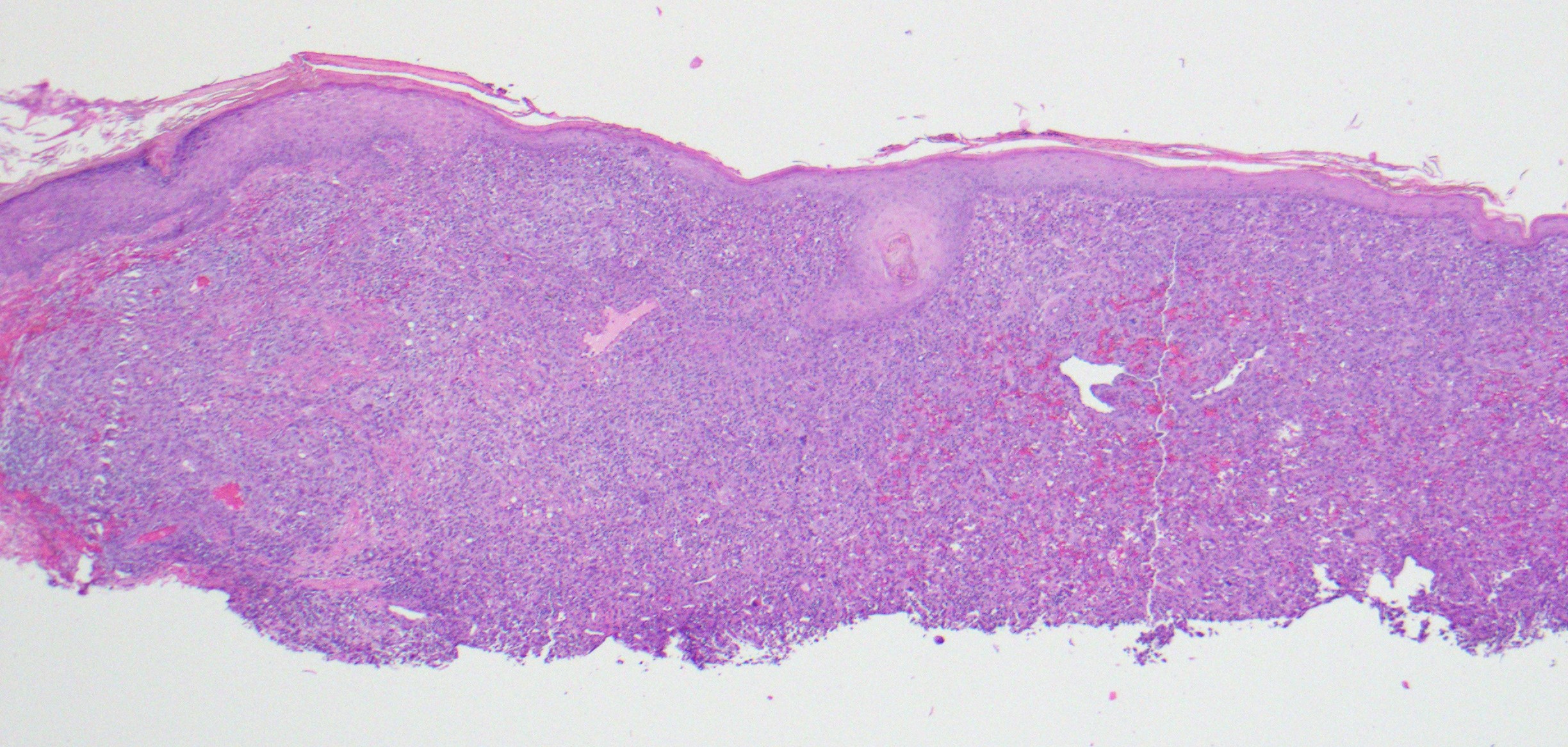
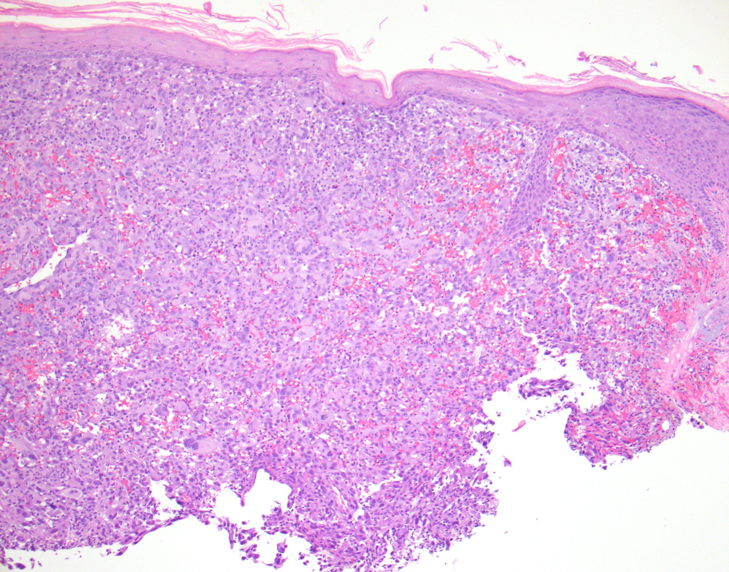
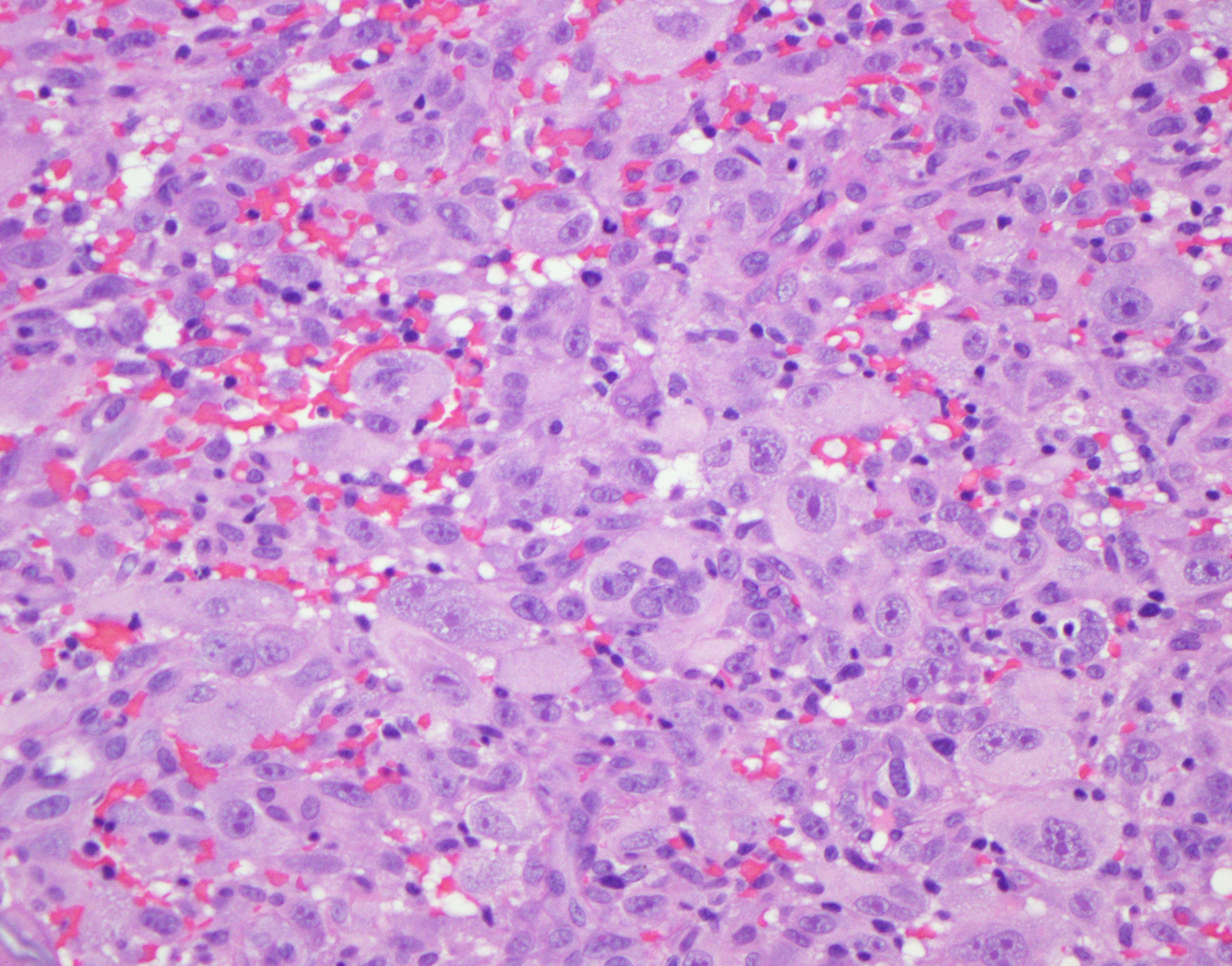
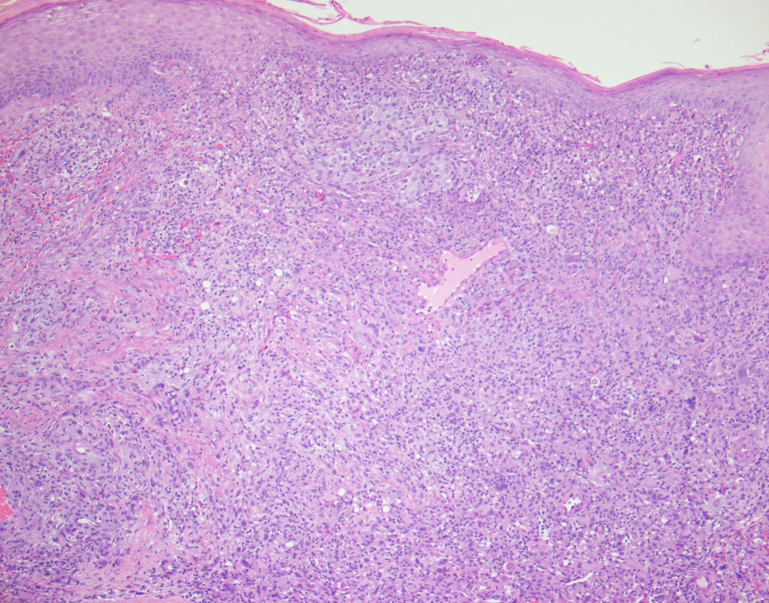
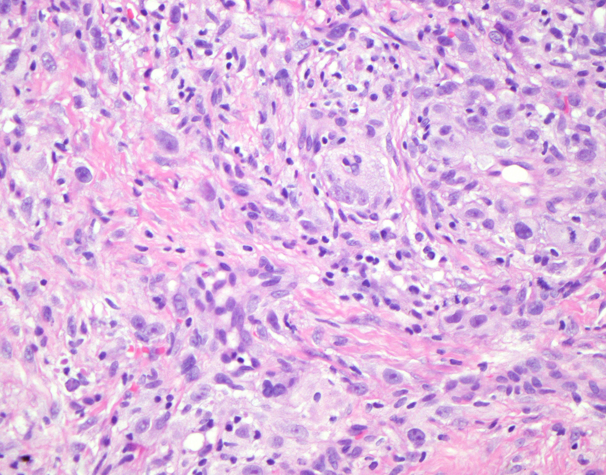
Final Diagnosis: Poorly Differentiated Epithelioid Malignancy Most Reminiscent of a Pleomorphic Dermal Sarcoma/Atypical Fibroxanthoma, Cannot Rule Out Dedifferentiated and Undifferentiated Melanoma with Pleomorphic Dermal Sarcoma Sarcomatoid Transdifferentiation.
Diagnosis comment: The tumor does not show any classic immunohistochemical profile that would support an epithelial, conventional melanocytic, vascular, neural, smooth muscle or hematopoietic line of differentiation. The diagnostic considerations are:
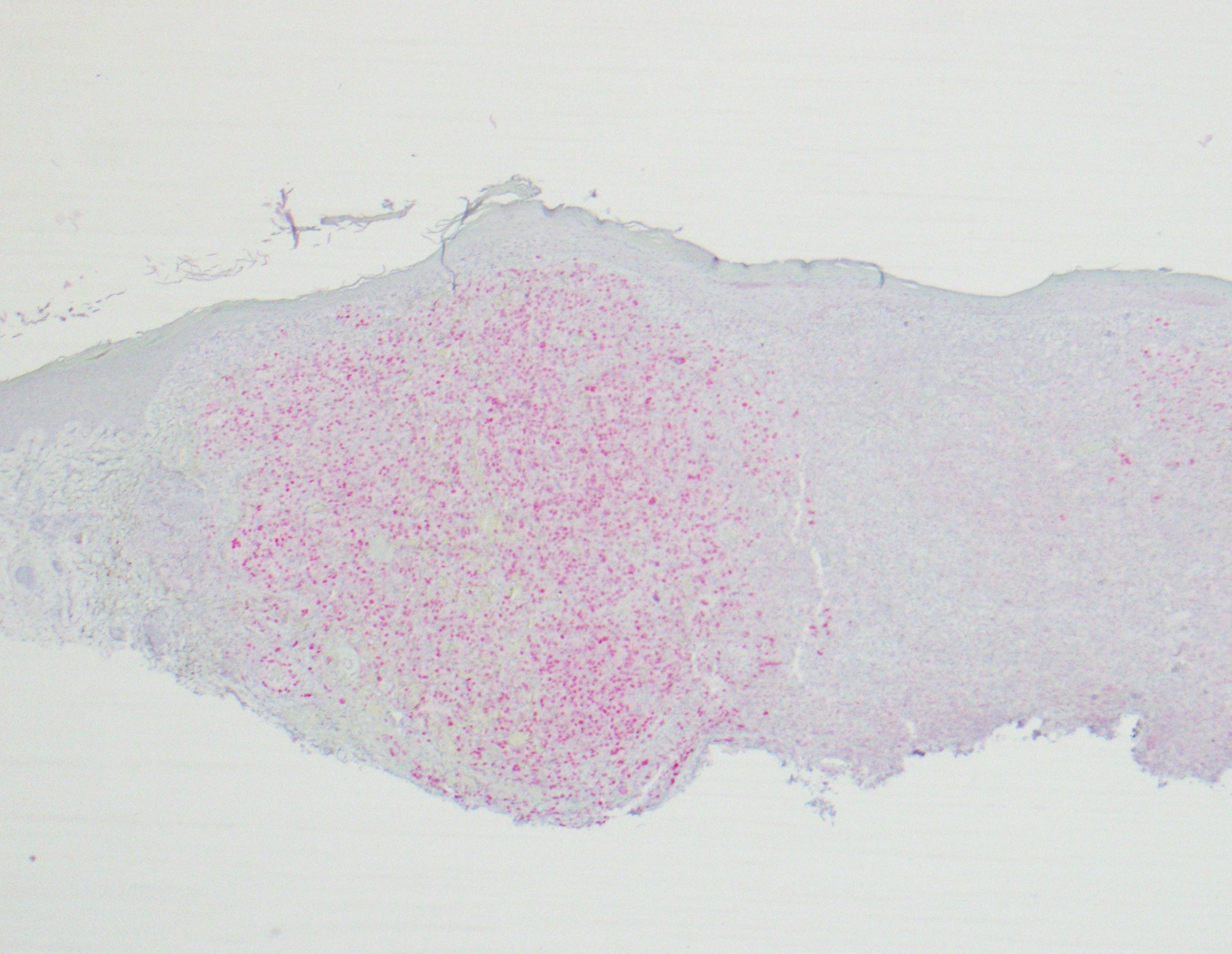
1. A pleomorphic dermal sarcoma/atypical fibroxanthoma that exhibits aberrant expression of microphthalmia transcription factor(minimal), PRAME and NK1/C3(see references).
2. A dedifferentiated or undifferentiated melanoma characterized by a tumor without positivity for conventional melanoma markers i.e. melan-A, HMB45, S100 and SOX10 and where the baseline histomorphology could be encountered in a sarcoma, perhaps best exemplified by a cutaneous equivalent of an undifferentiated pleomorphic sarcoma.
3. A collision of a pleomorphic dermal sarcoma/atypical fibroxanthoma and a dedifferentiated and undifferentiated melanoma, either representing a true collision tumor of both melanoma and pleomorphic dermal sarcoma or further sarcomatoid transdifferentiation in a dedifferentiated and undifferentiated melanoma.
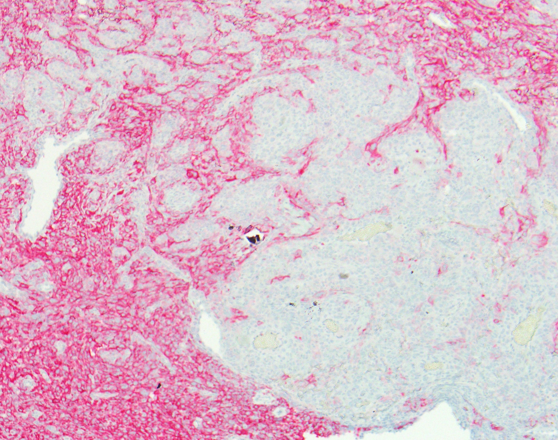
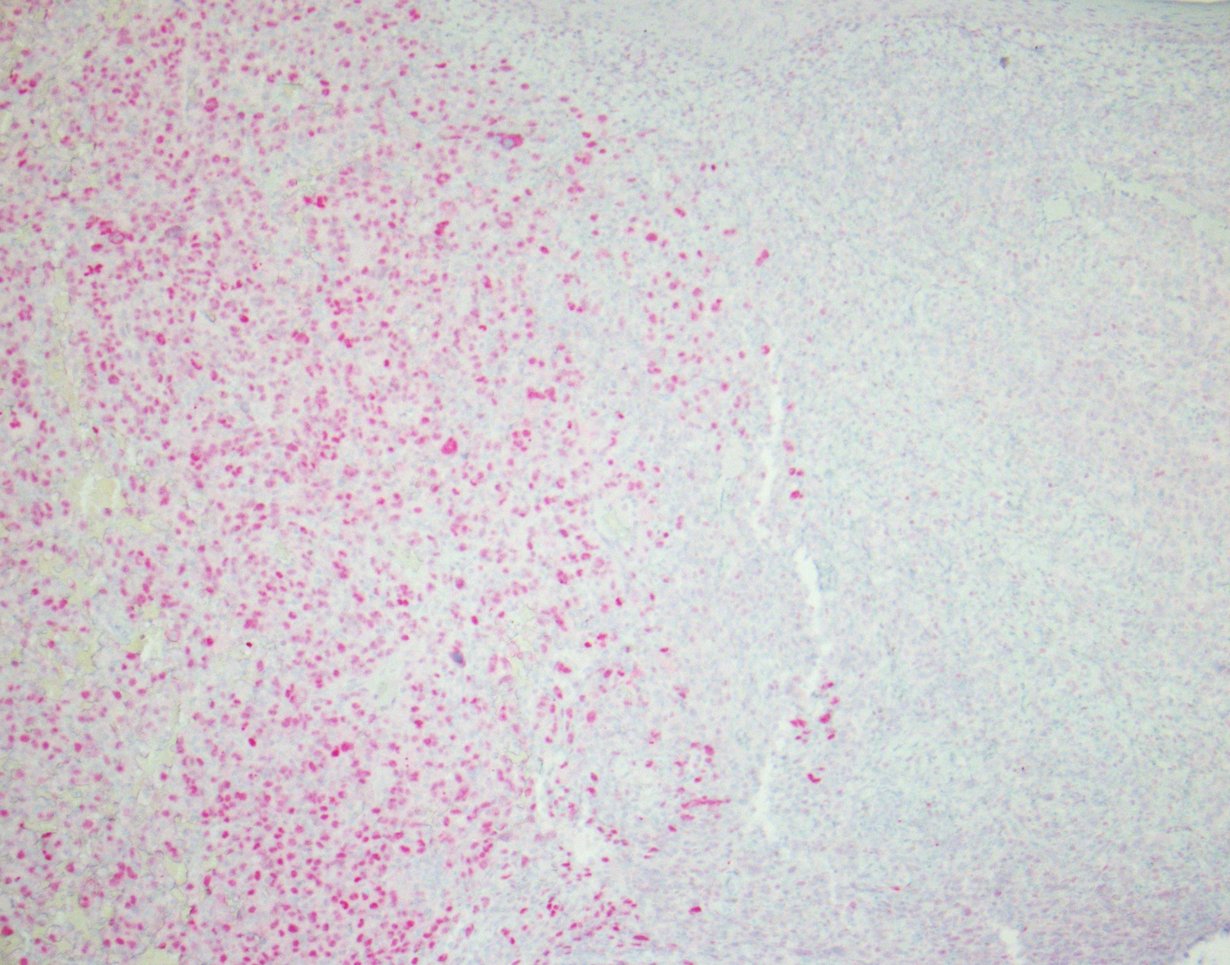
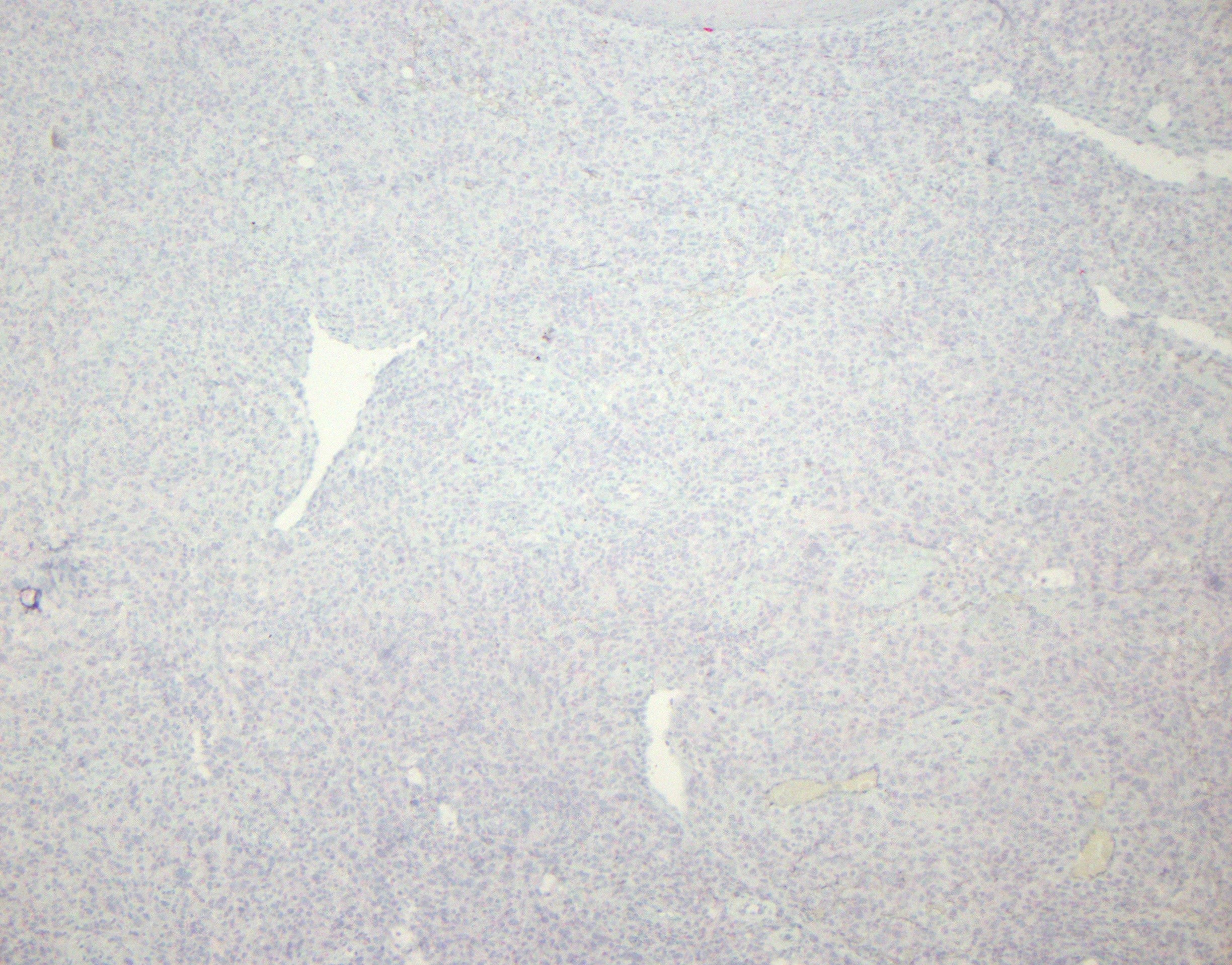
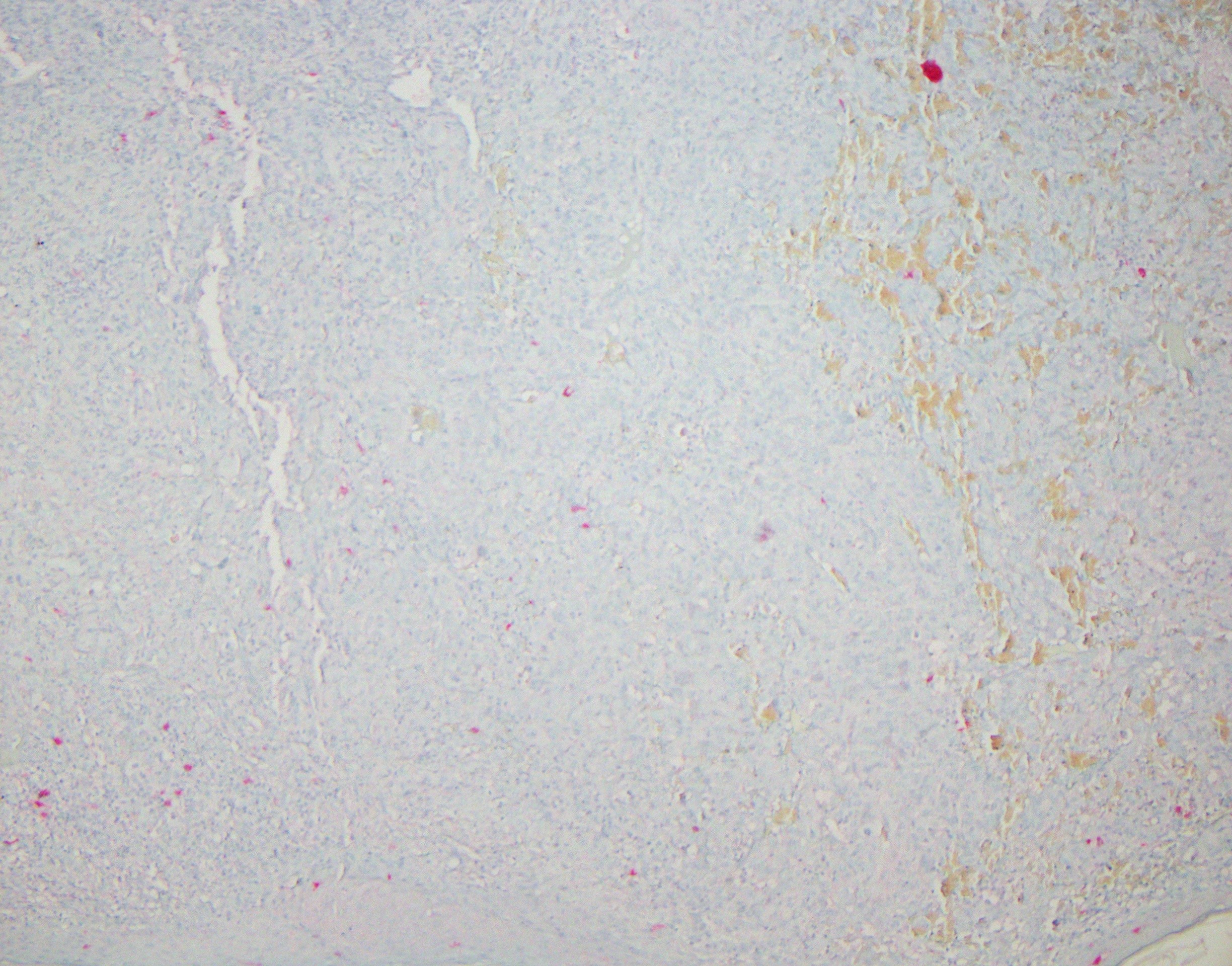
The CD10 staining pattern along with the PRAME staining pattern perhaps interesting. In particular, on the CD10 preparation, there is a distinct area where the tumor is not CD10 positive. CD10, as you know, is characteristically expressed in pleomorphic dermal sarcoma/atypical fibroxanthoma, despite the fact that it is not a very specific stain. It is also interesting to note that where the tumor is not expressing CD10 corresponds to significant area of pan nuclear staining for PRAME. I do not know if this difference in staining pattern is a clue to the potential melanocyte ontogeny of the tumor defining therefore a dedifferentiated melanoma or if the difference in staining compared to the remainder of the tumor simply reflects some element of heterogeneity in the molecular profile of the tumor perhaps analagous to the variable staining patterns that can be observed for S100 or melan-A in a melanoma.
Click images to begin the slideshow