Final diagnosis: Borderline Type-C Lymphomatoid Papulosis.
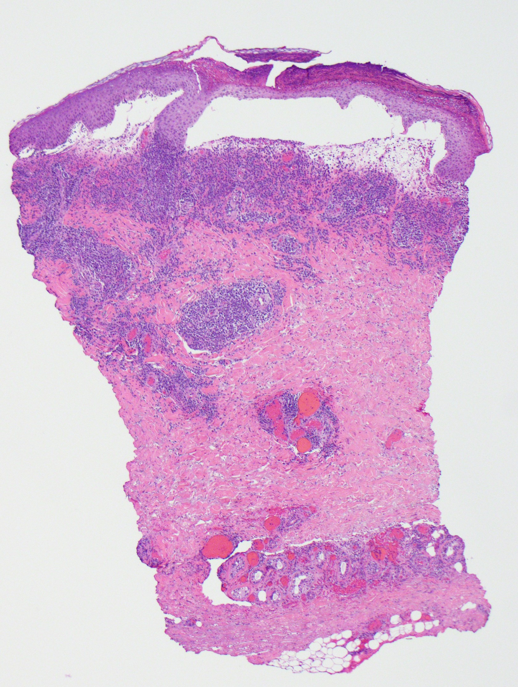
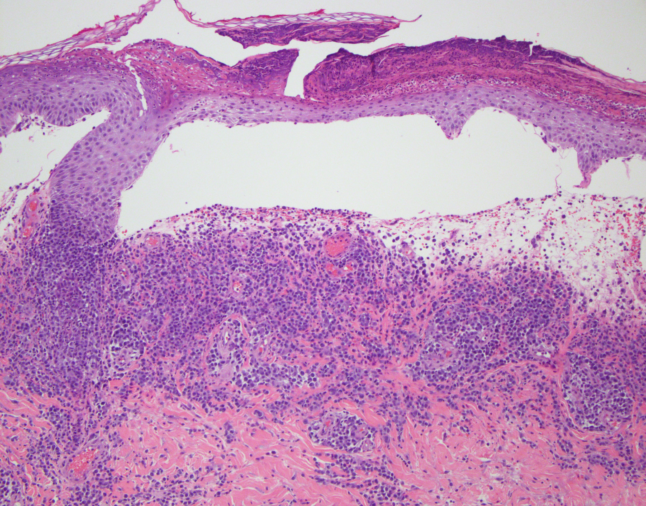
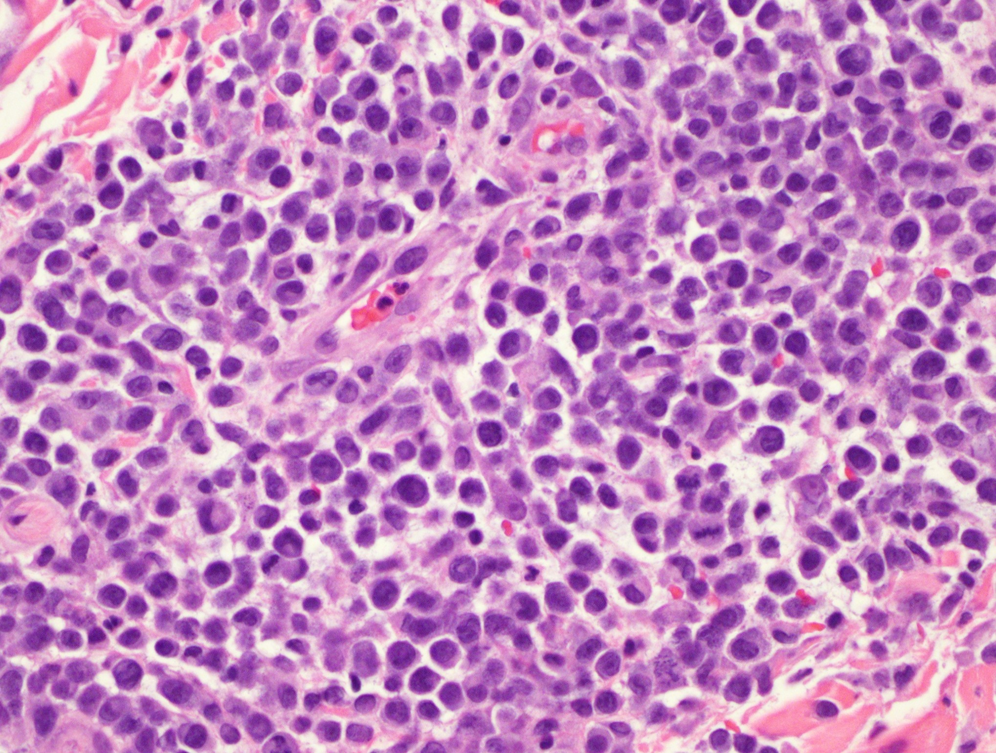
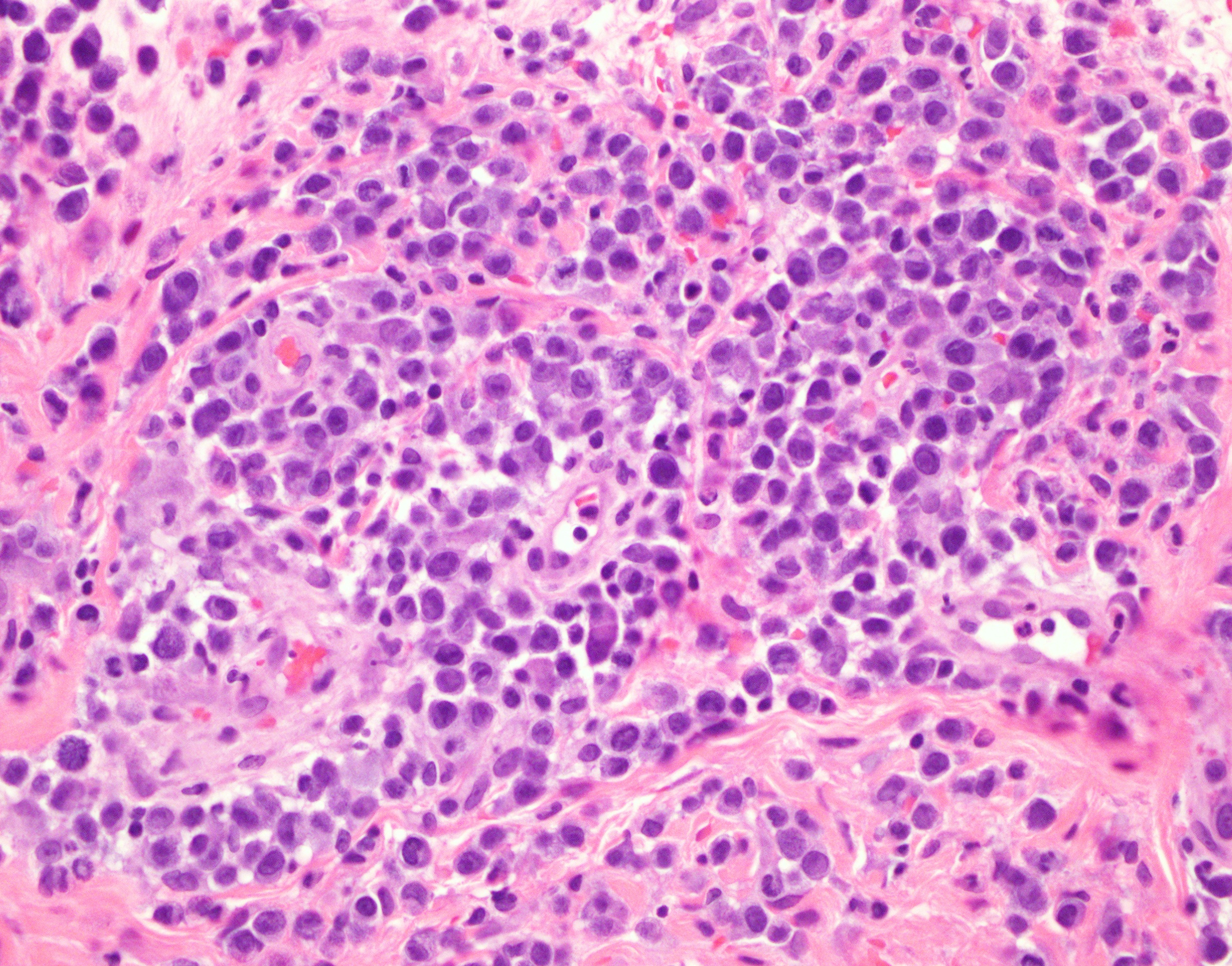
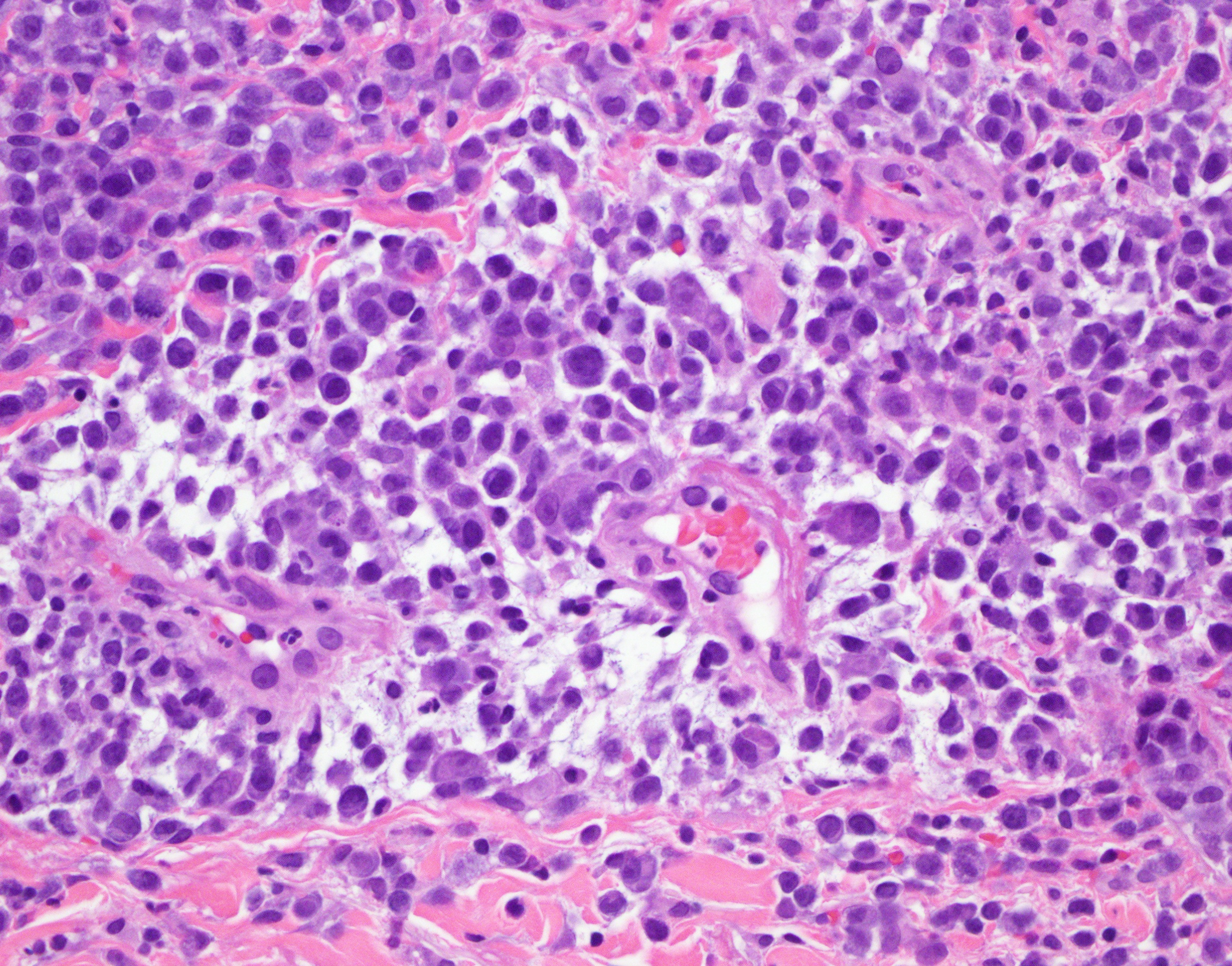
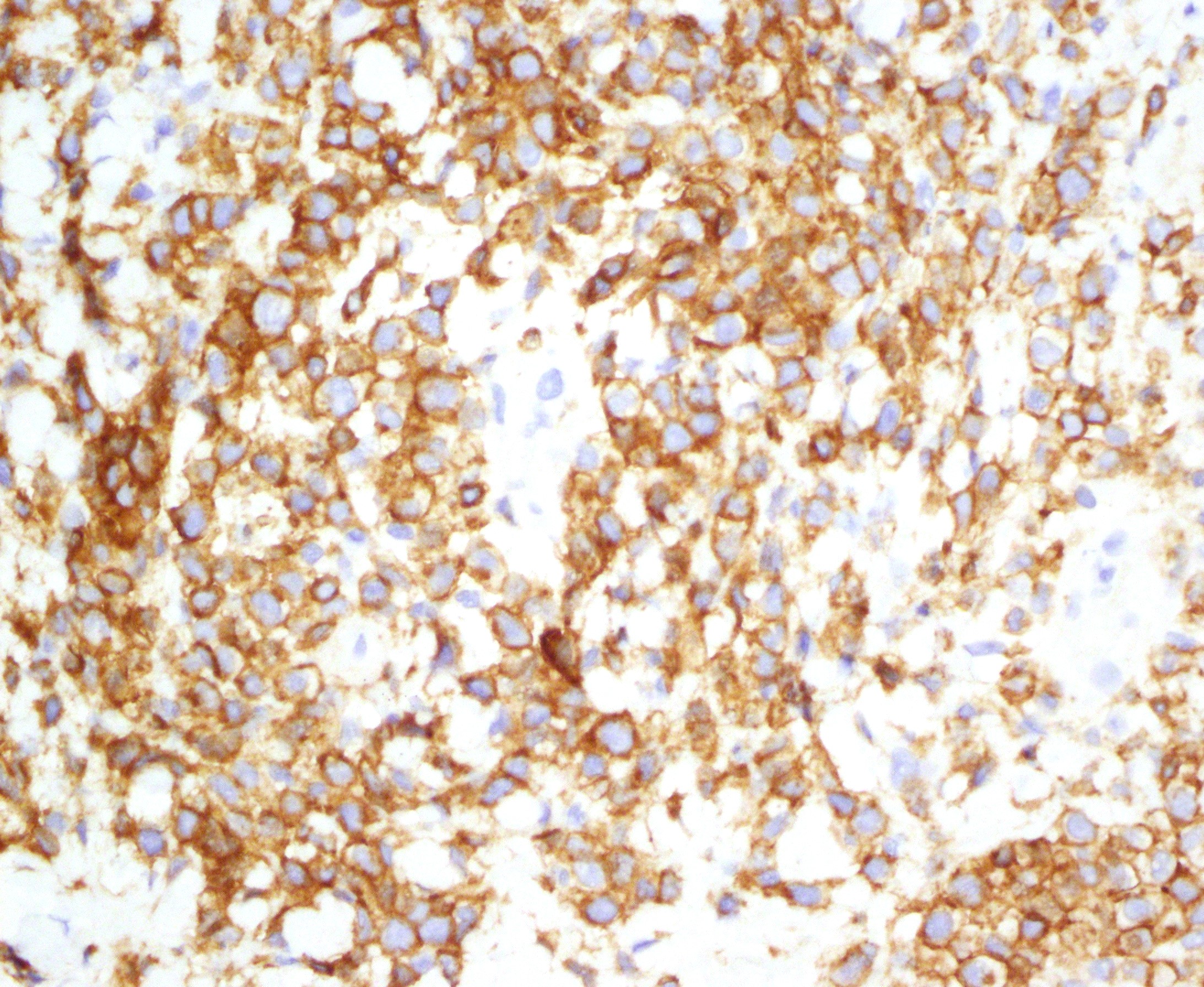
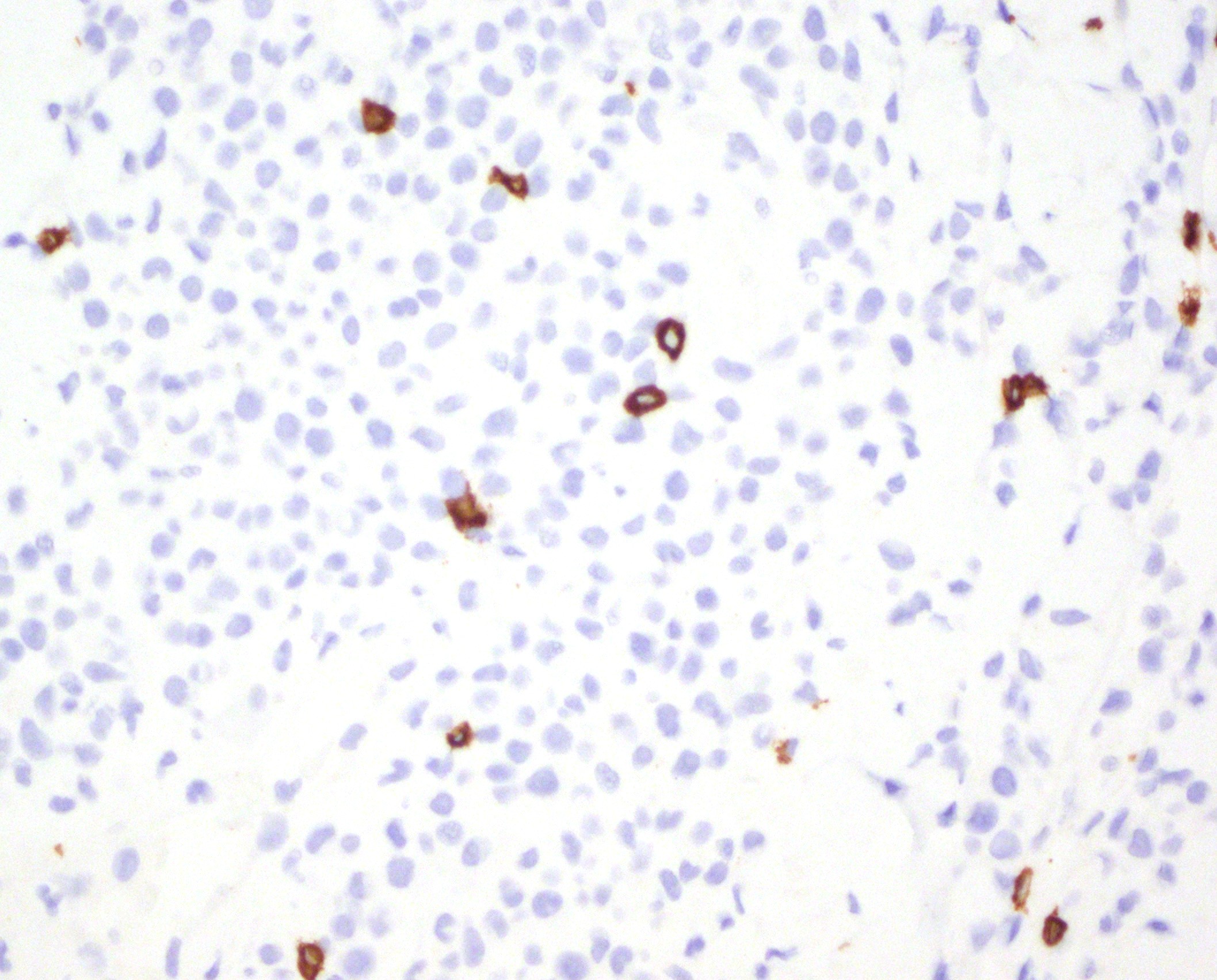
Diagnosis comment: The biopsy shows a highly atypical transformed neoplastic CD4-positive, CD30-positive angiocentric and superficial diffuse atypical lymphocytic infiltrate of the alpha beta subset. The CD4 positive T-cells also exhibit cytotoxic protein expression, given the degree of immunoreactivity for granzyme and TIA. Not surprisingly there is a variable reduction in staining for select pan T cell markers namely CD3, CD5, and CD7. The overlying epidermis exhibits a pustular psoriasiform diathesis.
Overall, the distinct clinical presentation, along with this histopathology and phenotypic profile is very characteristic for a lymphomatoid papulosis as a form of indolent CD30 positive lymphoproliferative disease. The extent of large cell infiltration would fall under the rubric of borderline type-C lymphomatoid papulosis. In theory, the lesions should undergo spontaneous regression without any therapeutic intervention.
Click images to begin the slideshow